Background: COVID-19 is characterised by respiratory symptoms, which deteriorate into respiratory failure in a substantial proportion of cases, requiring intensive care in up to a third of patients admitted to hospital. Analysis of the pathological features in the lung tissues of patients who have died with COVID-19 could help us to understand the disease pathogenesis and clinical outcomes.
Methods: We systematically analysed lung tissue samples from 38 patients who died from COVID-19 in two hospitals in northern Italy between Feb 29 and March 24, 2020. The most representative areas identified at macroscopic examination were selected, and tissue blocks (median seven, range five to nine) were taken from each lung and fixed in 10% buffered formalin for at least 48 h. Tissues were assessed with use of haematoxylin and eosin staining, immunohistochemical staining for inflammatory infiltrate and cellular components (including staining with antibodies against CD68, CD3, CD45, CD61, TTF1, p40, and Ki-67), and electron microscopy to identify virion localisation.
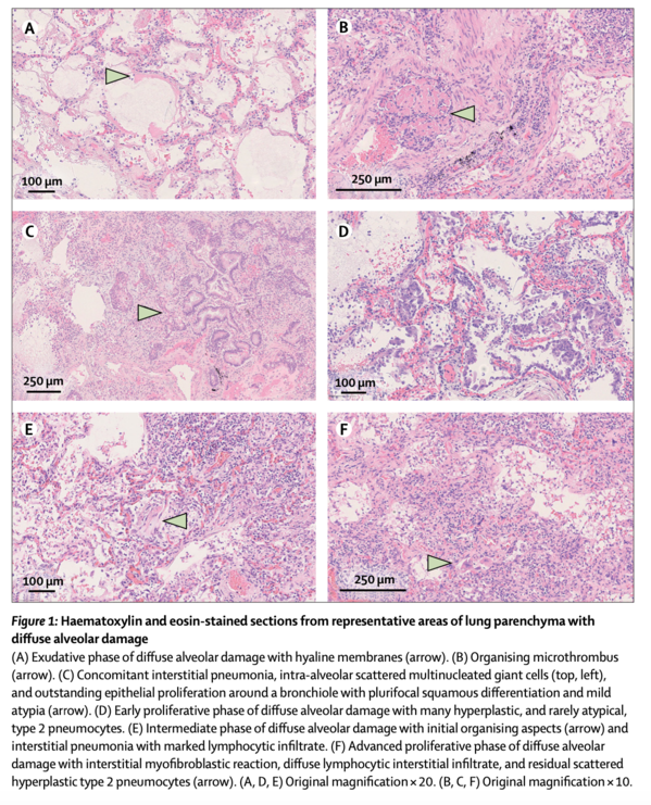
Findings: All cases showed features of the exudative and proliferative phases of diffuse alveolar damage, which included capillary congestion (in all cases), necrosis of pneumocytes (in all cases), hyaline membranes (in 33 cases), interstitial and intra-alveolar oedema (in 37 cases), type 2 pneumocyte hyperplasia (in all cases), squamous metaplasia with atypia (in 21 cases), and platelet-fibrin thrombi (in 33 cases). The inflammatory infiltrate, observed in all cases, was largely composed of macrophages in the alveolar lumina (in 24 cases) and lymphocytes in the interstitium (in 31 cases). Electron microscopy revealed that viral particles were predominantly located in the pneumocytes.
Interpretation: The predominant pattern of lung lesions in patients with COVID-19 patients is diffuse alveolar damage, as described in patients infected with severe acute respiratory syndrome and Middle East respiratory syndrome coronaviruses. Hyaline membrane formation and pneumocyte atypical hyperplasia are frequent. Importantly, the presence of platelet-fibrin thrombi in small arterial vessels is consistent with coagulopathy, which appears to be common in patients with COVID-19 and should be one of the main targets of therapy.
肺の剖検所見(北イタリア COVID-19ケースシリーズ):2施設の記述的研究(Lancet 2020.06.08)
(背景)
COVID-19は呼吸器症状を特徴とし、高い割合で呼吸不全を呈し、入院患者の3分の1が集中治療を必要とする。COVID-19で死亡した患者の肺組織の病理学的特徴を解析することで、疾患の病態と臨床転帰を理解することができる可能性がある。
(方法)
イタリア北部の2つの病院で2020年2月29日から3月24日までの間にCOVID-19で死亡した患者38人の肺組織サンプルを系統的に分析した。マクロで最も代表的な部位を選択し、各肺から組織ブロック(中央値7、範囲5~9)を採取し、10%緩衝ホルマリン中で少なくとも48時間固定した。組織は、ヘマトキシリンおよびエオシン染色、炎症性浸潤および細胞成分の免疫組織化学染色(CD68、CD3、CD45、CD61、TTF1、p40、およびKi-67に対する抗体による染色を含む)、およびウイルスの局在を同定するための電子顕微鏡を使用して評価した。
(所見)
全例にびまん性肺胞損傷の滲出・増殖期の特徴が認められ、毛細血管のうっ血(全例)、肺胞上皮細胞の壊死(全例)、肺硝子膜(33例)、間質性・肺胞内水腫(37例)、2型肺胞上皮細胞過形成(全例)、異型を伴う扁平上皮変成(21例)、血小板・フィブリン血栓(33例)などが認められた。すべての症例で観察された炎症性浸潤は、主に肺胞腔内腔のマクロファージ(24例)と間質のリンパ球(31例)で構成されていた。電子顕微鏡検査の結果、ウイルス粒子は主に肺胞細胞に存在していた。
(解釈)
COVID-19患者の肺病変の優勢なパターンは、重症急性呼吸器症候群や中東呼吸器症候群コロナウイルス感染患者で報告されているようなびまん性肺胞損傷である。肺硝子膜形成および肺胞上皮細胞の非定型過形成が頻発している。重要なことに、小動脈血管における血小板-フィブリン血栓の存在は、COVID-19患者によく見られる凝固障害と一致しており、治療の主要ターゲットの一つとすべきである。
https://www.thelancet.com/journals/laninf/article/PIIS1473-3099(20)30434-5/fulltext